PodcastsCienciasHemispherics

Hemispherics

Hemispherics
Hemispherics
Último episodio

100 episodios

  • Hemispherics

    #98: Entrevista a Lisandro Sanabria. Sueño, memoria y aprendizaje en neurorrehabilitación

    01/06/2026 | 1 h 43 min
    En este episodio conversamos con Lisandro Sanabria, kinesiólogo argentino especializado en neurorrehabilitación, sobre una pregunta tan sencilla como profunda: ¿qué papel juega el sueño en la recuperación neurológica?

    A partir de su trayectoria clínica e investigadora, nos adentramos en la relación entre sueño, memoria y aprendizaje motor. Hablamos de ondas lentas, spindles talamocorticales, ripples hipocampales, sueño REM, reconsolidación y de cómo el cerebro podría seguir “trabajando” después de una sesión de terapia.

    El hilo conductor de la conversación es su investigación sobre la distancia temporal entre la rehabilitación y el sueño nocturno en pacientes con daño cerebral adquirido. Comentamos los resultados de un estudio retrospectivo con 50 pacientes, las tendencias observadas en quienes dormían poco después de la sesión y, sobre todo, las muchas preguntas que todavía quedan abiertas: cronotipo, insomnio, siestas, medicación, horarios, registros objetivos del sueño y diseño de futuros estudios.

    Una conversación sobre lo que ocurre cuando termina la sesión: cómo se consolida lo aprendido, qué tareas podrían beneficiarse más del sueño y por qué quizá la rehabilitación del futuro no solo deba preguntarse cuánto y qué entrenamos, sino también cuándo lo hacemos.

    Podéis encontrar a Lisandro en:

    LinkedIn: https://www.linkedin.com/in/lisandro-sanabria-665b40160/
    Instagram: https://www.instagram.com/lisandrosanabriadorignac/

    Referencias del episodio:

    Caffaro, P., Forcato, C., Gallo, F., & Sanabria, L. (2024). Impacto de la distancia temporal entre la sesión de rehabilitación al sueño nocturno en la recuperación física en pacientes con daño cerebral adquirido [Comunicación científica / póster].

    Dudai, Y. (2002). *Memory from A to Z: Keywords, concepts, and beyond*. Oxford University Press.

    Griffa, G., Solano, A., Deleglise, A., De Pino, G., Jacobacci, F., & Della-Maggiore, V. (2025). Uncovering the role of the human hippocampus in procedural motor learning: Insights from implicit sensorimotor adaptation. The Journal of Neuroscience, 45(36), e2119242025. https://doi.org/10.1523/JNEUROSCI.2119-24.2025

    Siengsukon, C. F., & Boyd, L. A. (2009). Sleep enhances off-line spatial and temporal motor learning after stroke. Neurorehabilitation and Neural Repair, 23(4), 327–335. https://doi.org/10.1177/1545968308326631

    Solano, A., Lerner, G., Griffa, G., Deleglise, A., Caffaro, P., Riquelme, L., Perez-Chada, D., & Della-Maggiore, V. (2024). Sleep consolidation potentiates sensorimotor adaptation. The Journal of Neuroscience, 44(36), e0325242024. https://doi.org/10.1523/JNEUROSCI.0325-24.2024
  • Hemispherics

    #97: El daño axonal difuso en el TCE

    16/05/2026 | 36 min
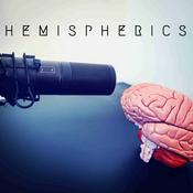
    En este episodio de Hemispherics hablamos sobre el daño axonal difuso tras un traumatismo craneoencefálico, una de las formas de lesión cerebral más frecuentes y, al mismo tiempo, más difíciles de comprender desde la clínica y la neuroimagen convencional. A lo largo del episodio revisamos cómo las fuerzas de aceleración y rotación pueden producir una lesión de desconexión en las redes cerebrales, profundizando en conceptos como la axotomía secundaria, la neuroinflamación, la vía del SARM1 o la lesión axonal traumática. También abordamos qué sabemos actualmente sobre resonancia magnética, tensor de difusión y biomarcadores como GFAP, UCH-L1 o neurofilamento ligero.

    Más allá de la biología, el episodio intenta trasladar todo esto a la realidad clínica y terapéutica. Hablamos de las expresiones cognitivas, conductuales y motoras que pueden aparecer en estos pacientes, de las limitaciones actuales del pronóstico y de cómo entender el daño axonal difuso no como una única lesión focal, sino como una alteración dinámica de redes cerebrales.

    Referencias del episodio:

    1. Adams, J. H., Doyle, D., Ford, I., Gennarelli, T. A., Graham, D. I., & McLellan, D. R. (1989). Diffuse axonal injury in head injury: definition, diagnosis and grading. Histopathology, 15(1), 49–59. https://doi.org/10.1111/j.1365-2559.1989.tb03040.x (https://pubmed.ncbi.nlm.nih.gov/2767623/).

    2. Bayley, M. T., Janzen, S., Harnett, A., Teasell, R., Patsakos, E., Marshall, S., Bragge, P., Velikonja, D., Kua, A., Douglas, J., Togher, L., Ponsford, J., & McIntyre, A. (2023). INCOG 2.0 Guidelines for Cognitive Rehabilitation Following Traumatic Brain Injury: Methods, Overview, and Principles. The Journal of head trauma rehabilitation, 38(1), 7–23. https://doi.org/10.1097/HTR.0000000000000838 (https://pubmed.ncbi.nlm.nih.gov/36594856/).

    3. Castaño-Leon, A. M., Sánchez Carabias, C., Hilario, A., Ramos, A., Navarro-Main, B., Paredes, I., Munarriz, P. M., Panero, I., Eiriz Fernández, C., García-Pérez, D., Moreno-Gomez, L. M., Esteban-Sinovas, O., Garcia Posadas, G., Gomez, P. A., & Lagares, A. (2022). Serum assessment of traumatic axonal injury: the correlation of GFAP, t-Tau, UCH-L1, and NfL levels with diffusion tensor imaging metrics and its prognosis utility. Journal of neurosurgery, 138(2), 454–464. https://doi.org/10.3171/2022.5.JNS22638 (https://pubmed.ncbi.nlm.nih.gov/35901687/).

    4. Frati, A., Cerretani, D., Fiaschi, A. I., Frati, P., Gatto, V., La Russa, R., Pesce, A., Pinchi, E., Santurro, A., Fraschetti, F., & Fineschi, V. (2017). Diffuse Axonal Injury and Oxidative Stress: A Comprehensive Review. International journal of molecular sciences, 18(12), 2600. https://doi.org/10.3390/ijms18122600 (https://pubmed.ncbi.nlm.nih.gov/29207487/).

    5. Geiger, P., Gmeiner, R., Schön, V., Petr, O., Thomé, C., & Pinggera, D. (2025). Timing of Magnetic Resonance Imaging (MRI) in Moderate and Severe TBI: A Systematic Review. Journal of clinical medicine, 14(12), 4078. https://doi.org/10.3390/jcm14124078 (https://pubmed.ncbi.nlm.nih.gov/40565823/).

    6. Henninger, N., Bouley, J., Sikoglu, E. M., An, J., Moore, C. M., King, J. A., Bowser, R., Freeman, M. R., & Brown, R. H., Jr (2016). Attenuated traumatic axonal injury and improved functional outcome after traumatic brain injury in mice lacking Sarm1. Brain : a journal of neurology, 139(Pt 4), 1094–1105. https://doi.org/10.1093/brain/aww001 (https://pubmed.ncbi.nlm.nih.gov/26912636/).

    7. Johnson, V. E., Stewart, W., & Smith, D. H. (2013). Axonal pathology in traumatic brain injury. Experimental neurology, 246, 35–43. https://doi.org/10.1016/j.expneurol.2012.01.013 (https://pubmed.ncbi.nlm.nih.gov/22285252/).

    8. Lagares, A., de la Cruz, J., Terrisse, H., Mejan, O., Pavlov, V., Vermorel, C., Payen, J. F., & of the BRAINI participants and investigators (2024). An automated blood test for glial fibrillary acidic protein (GFAP) and ubiquitin carboxy-terminal hydrolase L1 (UCH-L1) to predict the absence of intracranial lesions on head CT in adult patients with mild traumatic brain injury: BRAINI, a multicentre observational study in Europe. EBioMedicine, 110, 105477. https://doi.org/10.1016/j.ebiom.2024.105477 (https://pmc.ncbi.nlm.nih.gov/articles/PMC11647500/).

    9. Mac Donald, C. L., Dikranian, K., Song, S. K., Bayly, P. V., Holtzman, D. M., & Brody, D. L. (2007). Detection of traumatic axonal injury with diffusion tensor imaging in a mouse model of traumatic brain injury. Experimental neurology, 205(1), 116–131. https://doi.org/10.1016/j.expneurol.2007.01.035 (https://pubmed.ncbi.nlm.nih.gov/17368446/).

    10. Mac Donald, C. L., Yuh, E. L., Vande Vyvere, T., Edlow, B. L., Li, L. M., Mayer, A. R., Mukherjee, P., Newcombe, V. F. J., Wilde, E. A., Koerte, I. K., Yurgelun-Todd, D., Wu, Y. C., Duhaime, A. C., Awwad, H. O., Dams-O'Connor, K., Doperalski, A., Maas, A. I. R., McCrea, M. A., Umoh, N., & Manley, G. T. (2025). Neuroimaging Characterization of Acute Traumatic Brain Injury with Focus on Frontline Clinicians: Recommendations from the 2024 National Institute of Neurological Disorders and Stroke Traumatic Brain Injury Classification and Nomenclature Initiative Imaging Working Group. Journal of neurotrauma, 42(13-14), 1056–1064. https://doi.org/10.1089/neu.2025.0079 (https://pubmed.ncbi.nlm.nih.gov/40393517/).

    11. Muehlschlegel, S., Rajajee, V., Wartenberg, K. E., Alexander, S. A., Busl, K. M., Creutzfeldt, C. J., Fontaine, G. V., Hocker, S. E., Hwang, D. Y., Kim, K. S., Madzar, D., Mahanes, D., Mainali, S., Meixensberger, J., Sakowitz, O. W., Varelas, P. N., Weimar, C., & Westermaier, T. (2024). Guidelines for Neuroprognostication in Critically Ill Adults with Moderate-Severe Traumatic Brain Injury. Neurocritical care, 40(2), 448–476. https://doi.org/10.1007/s12028-023-01902-2 (https://pubmed.ncbi.nlm.nih.gov/38366277/).

    12. Ponsford, J. L., Downing, M. G., Olver, J., Ponsford, M., Acher, R., Carty, M., & Spitz, G. (2014). Longitudinal follow-up of patients with traumatic brain injury: outcome at two, five, and ten years post-injury. Journal of neurotrauma, 31(1), 64–77. https://doi.org/10.1089/neu.2013.2997 (https://pubmed.ncbi.nlm.nih.gov/23889321/).

    13. Sassani, M., Ghafari, T., Arachchige, P. R. W., Idrees, I., Gao, Y., Waitt, A., Weaver, S. R. C., Mazaheri, A., Lyons, H. S., Grech, O., Thaller, M., Witton, C., Bagshaw, A. P., Wilson, M., Park, H., Brookes, M., Novak, J., Mollan, S. P., Hill, L. J., Lucas, S. J. E., … Fernández-Espejo, D. (2025). Current and prospective roles of magnetic resonance imaging in mild traumatic brain injury. Brain communications, 7(2), fcaf120. https://doi.org/10.1093/braincomms/fcaf120 (https://pubmed.ncbi.nlm.nih.gov/40241788/).

    14. Siedler, D. G., Chuah, M. I., Kirkcaldie, M. T., Vickers, J. C., & King, A. E. (2014). Diffuse axonal injury in brain trauma: insights from alterations in neurofilaments. Frontiers in cellular neuroscience, 8, 429. https://doi.org/10.3389/fncel.2014.00429 (https://pubmed.ncbi.nlm.nih.gov/25565963/).

    15. Smith, D. H., Hicks, R., & Povlishock, J. T. (2013). Therapy development for diffuse axonal injury. Journal of neurotrauma, 30(5), 307–323. https://doi.org/10.1089/neu.2012.2825 (https://pubmed.ncbi.nlm.nih.gov/23252624/).

    16. Wofford, K. L., Loane, D. J., & Cullen, D. K. (2019). Acute drivers of neuroinflammation in traumatic brain injury. Neural regeneration research, 14(9), 1481–1489. https://doi.org/10.4103/1673-5374.255958 (https://pmc.ncbi.nlm.nih.gov/articles/PMC6557091/).
  • Hemispherics

    #96: La mano 'alien' tras un daño cerebral

    02/05/2026 | 45 min
    La mano alien es uno de esos fenómenos neurológicos que obligan a replantearse qué significa realmente “controlar” una acción: pacientes cuya mano no está paralizada, pero tampoco les obedece, realizando movimientos con apariencia intencional que surgen fuera de su voluntad e incluso interfieren con la otra mano. En este episodio utilizamos este cuadro tan llamativo como clínicamente revelador para ir mucho más allá del síntoma y explorar cómo el cerebro construye la acción, integrando intención, ejecución y percepción dentro de una red compleja que, cuando se desorganiza, rompe la coherencia entre lo que queremos hacer y lo que finalmente ocurre. Desgranamos los distintos fenotipos —frontal, calloso y parietal— como expresiones de fallos en nodos específicos de esa red, analizamos su base neurofisiológica y aterrizamos todo esto en la clínica. Cómo reconocer la mano alien, cómo valorarla desde la fenomenología y la interacción con el entorno, y qué estrategias terapéuticas pueden tener sentido en función del mecanismo predominante. Un episodio que no solo explica un síndrome raro, sino que abre una ventana para entender que el movimiento no es simplemente contraer músculos, sino construir continuamente la experiencia de ser quien actúa.

    Referencias del episodio:

    1.     Biran, I., Giovannetti, T., Buxbaum, L., & Chatterjee, A. (2006). The alien hand syndrome: What makes the alien hand alien?. Cognitive neuropsychology, 23(4), 563–582. https://doi.org/10.1080/02643290500180282 (https://pubmed.ncbi.nlm.nih.gov/21049344/).

    2.     Bru, I., Verhamme, L., de Neve, P., & Maebe, H. (2021). Rehabilitation of a Patient with Alien Hand Syndrome: a Case Report of a 61-Year Old Man. Journal of rehabilitation medicine. Clinical communications, 4, 1000050. https://doi.org/10.2340/20030711-1000050 (https://pmc.ncbi.nlm.nih.gov/articles/PMC8054745/).

    3.     Di Pietro, M., Russo, M., Dono, F., Carrarini, C., Thomas, A., Di Stefano, V., Telese, R., Bonanni, L., Sensi, S. L., Onofrj, M., & Franciotti, R. (2021). A Critical Review of Alien Limb-Related Phenomena and Implications for Functional Magnetic Resonance Imaging Studies. Frontiers in neurology, 12, 661130. https://doi.org/10.3389/fneur.2021.661130 (https://pmc.ncbi.nlm.nih.gov/articles/PMC8458742/).

    4.     Feinberg, T. E., Schindler, R. J., Flanagan, N. G., & Haber, L. D. (1992). Two alien hand syndromes. Neurology, 42(1), 19–24. https://doi.org/10.1212/wnl.42.1.19 (https://pubmed.ncbi.nlm.nih.gov/1734302/).

    5.     Graff-Radford, J., Rubin, M. N., Jones, D. T., Aksamit, A. J., Ahlskog, J. E., Knopman, D. S., Petersen, R. C., Boeve, B. F., & Josephs, K. A. (2013). The alien limb phenomenon. Journal of neurology, 260(7), 1880–1888. https://doi.org/10.1007/s00415-013-6898-y (https://pubmed.ncbi.nlm.nih.gov/23572346/).

    6.     Haq, I. U., Malaty, I. A., Okun, M. S., Jacobson, C. E., Fernandez, H. H., & Rodriguez, R. R. (2010). Clonazepam and botulinum toxin for the treatment of alien limb phenomenon. The neurologist, 16(2), 106–108. https://doi.org/10.1097/NRL.0b013e3181a0d670 (https://pubmed.ncbi.nlm.nih.gov/20220444/).

    7.     Hassan, A., & Josephs, K. A. (2016). Alien Hand Syndrome. Current neurology and neuroscience reports, 16(8), 73. https://doi.org/10.1007/s11910-016-0676-z (https://pubmed.ncbi.nlm.nih.gov/27315251/).

    8.     Lewis-Smith, D. J., Wolpe, N., Ghosh, B. C. P., & Rowe, J. B. (2020). Alien limb in the corticobasal syndrome: phenomenological characteristics and relationship to apraxia. Journal of neurology, 267(4), 1147–1157. https://doi.org/10.1007/s00415-019-09672-8 (https://pmc.ncbi.nlm.nih.gov/articles/PMC7109196/).

    9.     Ma, Y., Liu, Y., Yan, X., & Ouyang, Y. (2023). Alien hand syndrome, a rare presentation of corpus callosum and cingulate infarction. Journal of the neurological sciences, 452, 120739. https://doi.org/10.1016/j.jns.2023.120739 (https://pubmed.ncbi.nlm.nih.gov/37536055/).

    10.  Mark V. W. (2025). Alien Hand: Current Research Trends. Current neurology and neuroscience reports, 25(1), 63. https://doi.org/10.1007/s11910-025-01449-z (https://pmc.ncbi.nlm.nih.gov/articles/PMC12449344/).

    11.  Park, Y. W., Kim, C. H., Kim, M. O., Jeong, H. J., & Jung, H. Y. (2012). Alien hand syndrome in stroke - case report & neurophysiologic study -. Annals of rehabilitation medicine, 36(4), 556–560. https://doi.org/10.5535/arm.2012.36.4.556 (https://pmc.ncbi.nlm.nih.gov/articles/PMC3438424/).

    12.  Romano, D., Sedda, A., Dell'aquila, R., Dalla Costa, D., Beretta, G., Maravita, A., & Bottini, G. (2014). Controlling the alien hand through the mirror box. A single case study of alien hand syndrome. Neurocase, 20(3), 307–316. https://doi.org/10.1080/13554794.2013.770882 (https://pubmed.ncbi.nlm.nih.gov/23557374/).

    13.  Sarva, H., Deik, A., & Severt, W. L. (2014). Pathophysiology and treatment of alien hand syndrome. Tremor and other hyperkinetic movements (New York, N.Y.), 4, 241. https://doi.org/10.7916/D8VX0F48 (https://pmc.ncbi.nlm.nih.gov/articles/PMC4261226/).

    14.  Sellal, F., Cretin, B., Musacchio, M., Berthel, M. C., Carelli, G., & Michel, J. M. (2019). Long-lasting diagonistic dyspraxia suppressed by rTMS applied to the right motor cortex. Journal of neurology, 266(3), 631–635. https://doi.org/10.1007/s00415-018-09178-9 (https://pubmed.ncbi.nlm.nih.gov/30631917/).

    15.  Wolpe, N., Moore, J. W., Rae, C. L., Rittman, T., Altena, E., Haggard, P., & Rowe, J. B. (2014). The medial frontal-prefrontal network for altered awareness and control of action in corticobasal syndrome. Brain : a journal of neurology, 137(Pt 1), 208–220. https://doi.org/10.1093/brain/awt302 (https://pmc.ncbi.nlm.nih.gov/articles/PMC3891444/).

    16.  Wolpe, N., Hezemans, F. H., & Rowe, J. B. (2020). Alien limb syndrome: A Bayesian account of unwanted actions. Cortex; a journal devoted to the study of the nervous system and behavior, 127, 29–41. https://doi.org/10.1016/j.cortex.2020.02.002 (https://pubmed.ncbi.nlm.nih.gov/32155475/).
  • Hemispherics

    #95: Afasia bilingüe. Cuando el cerebro olvida un idioma

    18/04/2026 | 54 min
    En este episodio nos metemos en un fenómeno tan curioso como clínicamente muy revelador: qué pasa cuando una persona bilingüe sufre un daño cerebral y, de repente, uno de sus idiomas parece desaparecer mientras el otro se mantiene. A partir de ahí, aprovechamos para repensar cómo se organiza realmente el lenguaje en el cerebro, alejándonos de la idea clásica de “zonas” y acercándonos a una visión mucho más dinámica, basada en redes que interactúan constantemente. Hablamos de por qué no todos los idiomas se afectan igual, qué papel tienen factores como el uso previo o la edad de adquisición, y por qué la recuperación puede ser tan variable entre pacientes. También aterrizamos todo esto en la clínica, viendo qué implicaciones tiene para la neurorrehabilitación y cómo trabajar con pacientes bilingües. En el fondo, es un episodio que va más allá de la afasia bilingüe y que nos ayuda a entender algo más profundo: que el lenguaje no está guardado en compartimentos, sino que es un sistema flexible que el cerebro reorganiza cuando algo se rompe.

    Referencias del episodio:

    1. Abutalebi, J., & Green, D. W. (2007). Bilingual language production: The neurocognition of language representation and control. Journal of Neurolinguistics, 20(3), 242–275.
    https://doi.org/10.1016/j.jneuroling.2006.10.003

    2. Abutalebi, J., Della Rosa, P. A., Gonzaga, A. K., Keim, R., Costa, A., & Perani, D. (2013). The role of the left putamen in multilingual language production. Brain and language, 125(3), 307–315. https://doi.org/10.1016/j.bandl.2012.03.009 (https://pubmed.ncbi.nlm.nih.gov/22538086/).

    3. Abutalebi, J., Canini, M., Della Rosa, P. A., Green, D. W., & Weekes, B. S. (2015). The neuroprotective effects of bilingualism upon the inferior parietal lobule: A structural neuroimaging study in aging Chinese bilinguals. Journal of Neurolinguistics, 26(6), 602–609 (https://www.sciencedirect.com/science/article/pii/S0911604414000657).

    4. Ardila, A., Lahiri, D., & Mukherjee, A. (2023). Bilingualism as a protective factor in aphasia. Applied neuropsychology. Adult, 30(5), 512–520. https://doi.org/10.1080/23279095.2021.1960837 (https://pubmed.ncbi.nlm.nih.gov/34392763/).

    5. Berthier, M. L. (2005). Poststroke aphasia: Epidemiology, pathophysiology and treatment. Drugs & Aging, 22(2), 163–182.
    https://doi.org/10.2165/00002512-200522020-00006 (https://pubmed.ncbi.nlm.nih.gov/15733022/).

    6. Bihovsky, A., Ben-Shachar, M., & Meir, N. (2024). Semantic feature analysis (SFA) treatment in L1 and L2 in bilingual aphasia: Effects of cognitive and language factors. Aphasiology, 38(4), 683–711 (https://www.tandfonline.com/doi/full/10.1080/02687038.2023.2226359).

    7. Dekhtyar, M., Kiran, S., & Gray, T. (2020). Is bilingualism protective for adults with aphasia?. Neuropsychologia, 139, 107355. https://doi.org/10.1016/j.neuropsychologia.2020.107355 (https://pubmed.ncbi.nlm.nih.gov/31972233/).

    8. Edmonds, L. A., & Kiran, S. (2006). Effect of semantic naming treatment on crosslinguistic generalization in bilingual aphasia. Journal of speech, language, and hearing research : JSLHR, 49(4), 729–748. https://doi.org/10.1044/1092-4388(2006/053) (https://pubmed.ncbi.nlm.nih.gov/16908872/).

    9. Goral, M., Levy, E. S., & Kastl, R. (2007). Cross-language treatment generalisation: A case of trilingual aphasia. Aphasiology, 103(1-2), 203–204. https://doi.org/10.1016/j.bandl.2007.07.116 (https://pubmed.ncbi.nlm.nih.gov/20221311/).

    10. Hickok, G., & Poeppel, D. (2007). The cortical organization of speech processing. Nature reviews. Neuroscience, 8(5), 393–402. https://doi.org/10.1038/nrn2113 (https://pubmed.ncbi.nlm.nih.gov/17431404/).

    11. Indefrey, P. (2006). A meta-analysis of hemodynamic studies on first and second language processing. Human Brain Mapping, 27(10), 799–810.
    https://doi.org/10.1002/hbm.20250 (https://onlinelibrary.wiley.com/doi/abs/10.1111/j.1467-9922.2006.00365.x).

    12. Jouravlev, O., Mineroff, Z., Blank, I. A., & Fedorenko, E. (2021). The Small and Efficient Language Network of Polyglots and Hyper-polyglots. Cerebral cortex (New York, N.Y. : 1991), 31(1), 62–76. https://doi.org/10.1093/cercor/bhaa205 (https://pubmed.ncbi.nlm.nih.gov/32820332/).

    13. Malik-Moraleda, S., Ayyash, D., Gallée, J., Affourtit, J., Hoffmann, M., Mineroff, Z., Jouravlev, O., & Fedorenko, E. (2022). An investigation across 45 languages and 12 language families reveals a universal language network. Nature neuroscience, 25(8), 1014–1019. https://doi.org/10.1038/s41593-022-01114-5 (https://pubmed.ncbi.nlm.nih.gov/35856094/).

    14. Malik-Moraleda, S., Jouravlev, O., Taliaferro, M., Mineroff, Z., Cucu, T., Mahowald, K., Blank, I. A., & Fedorenko, E. (2024). Functional characterization of the language network of polyglots and hyperpolyglots with precision fMRI. bioRxiv : the preprint server for biology, 2023.01.19.524657. https://doi.org/10.1101/2023.01.19.524657 (https://pubmed.ncbi.nlm.nih.gov/36711949/).

    15. Nadeau, S. E. (2019). Neural basis of language: A population encoding perspective. Language, Cognition and Neuroscience, 34(1), 1–18.
    https://doi.org/10.1080/23273798.2018.1506034

    16. Paplikar, A., Mekala, S., Bak, T. H., Dharamkar, S., Alladi, S., & Kaul, S. (2018).
    Bilingualism and the severity of poststroke aphasia. Aphasiology, 33(1), 58–72. https://doi.org/10.1080/02687038.2017.1423272

    17. Pliatsikas, C. (2020). Understanding structural plasticity in the bilingual brain: The Dynamic Restructuring Model. Bilingualism: Language and Cognition, 23(2), 459–471. https://doi.org/10.1017/S1366728919000130

    18. Price C. J. (2012). A review and synthesis of the first 20 years of PET and fMRI studies of heard speech, spoken language and reading. NeuroImage, 62(2), 816–847. https://doi.org/10.1016/j.neuroimage.2012.04.062

    19. Russell-Meill, M., Marte, M. J., Carpenter, E., & Kiran, S. (2025). Navigating the Complexity of Bilingual Aphasia: Current Insights and Future Directions. Brain sciences, 15(9), 989. https://doi.org/10.3390/brainsci15090989 (https://pubmed.ncbi.nlm.nih.gov/41008350/).
  • Hemispherics

    #94: La sinergia flexora...¿una cárcel espinal?

    04/04/2026 | 34 min
    En este episodio de Hemispherics nos metemos de lleno en una de las preguntas clásicas de la neurorrehabilitación: ¿de dónde sale realmente la sinergia flexora? A partir de trabajos muy recientes con participación de Krakauer, exploramos una idea que puede cambiar la forma en la que entendemos este fenómeno: y es que quizá la sinergia no sea algo que el cerebro “genere” tras la lesión, sino la expresión de patrones preexistentes en la médula espinal que emergen cuando se pierde suficiente control descendente. A lo largo del episodio desmontamos el modelo clásico centrado en la vía reticuloespinal, analizamos evidencia experimental en primates y proponemos una visión integradora donde corteza, tronco y médula interactúan en distintos niveles. Un episodio para replantearse no solo el origen de la sinergia, sino también cómo la abordamos en clínica.

    Referencias del episodio:

    1. Baines, A., Poll, A., Baker, A. M., Krakauer, J. W., & Baker, S. N. (2026). Arm Control and its Recovery after Selective Lesions of Sensorimotor Cortex and the Red Nucleus: A Kinematic Study in Non-Human Primates. bioRxiv : the preprint server for biology, 2025.08.06.668715. https://doi.org/10.1101/2025.08.06.668715

    2. Baines, A., Glover, I. S., Baker, A. M. E., Krakauer, J. W., & Baker, S. N. (2026). Extent of damage to descending output from cortex rather than to specific cortical regions drives the emergence of flexor synergy in non-human primates. bioRxiv : the preprint server for biology, 2026.03.04.709517. https://doi.org/10.64898/2026.03.04.709517

    3. Brunnstrom, S. (1970). Movement therapy in hemiplegia: A neurophysiological approach. Harper & Row.

    4. Dewald, J. P., Pope, P. S., Given, J. D., Buchanan, T. S., & Rymer, W. Z. (1995). Abnormal muscle coactivation patterns during isometric torque generation at the elbow and shoulder in hemiparetic subjects. Brain : a journal of neurology, 118 ( Pt 2), 495–510. https://doi.org/10.1093/brain/118.2.495 (https://pubmed.ncbi.nlm.nih.gov/7735890/).

    5. Dewald, J. P., & Beer, R. F. (2001). Abnormal joint torque patterns in the paretic upper limb of subjects with hemiparesis. Muscle & nerve, 24(2), 273–283. https://doi.org/10.1002/1097-4598(200102)24:23.0.co;2-z (https://pubmed.ncbi.nlm.nih.gov/11180211/).

    6. Hug, F., Avrillon, S., Ibáñez, J., & Farina, D. (2023). Common synaptic input, synergies and size principle: Control of spinal motor neurons for movement generation. The Journal of physiology, 601(1), 11–20. https://doi.org/10.1113/JP283698 (https://pmc.ncbi.nlm.nih.gov/articles/PMC10098498/).

    7. Glover, I. S., Baker, A. M. E., Krakauer, J. W., & Baker, S. N. (2026). A spinal origin for the obligate flexor synergy in the non-human primate: Implications for control of reaching. The Journal of neuroscience : the official journal of the Society for Neuroscience, e1878252026. Advance online publication. https://doi.org/10.1523/JNEUROSCI.1878-25.2026 (https://pubmed.ncbi.nlm.nih.gov/41802867/).

    8. Kiehn O. (2016). Decoding the organization of spinal circuits that control locomotion. Nature reviews. Neuroscience, 17(4), 224–238. https://doi.org/10.1038/nrn.2016.9 (https://pubmed.ncbi.nlm.nih.gov/26935168/).

    9. McPherson, J. G., Chen, A., Ellis, M. D., Yao, J., Heckman, C. J., & Dewald, J. P. A. (2018). Progressive recruitment of contralesional cortico-reticulospinal pathways drives motor impairment post stroke. The Journal of physiology, 596(7), 1211–1225. https://doi.org/10.1113/JP274968 (https://pubmed.ncbi.nlm.nih.gov/29457651/).

    10. McPherson, L. M., & Dewald, J. P. A. (2022). Abnormal synergies and associated reactions post-hemiparetic stroke reflect muscle activation patterns of brainstem motor pathways. Frontiers in neurology, 13, 934670. https://doi.org/10.3389/fneur.2022.934670 (https://pubmed.ncbi.nlm.nih.gov/36299276/).

    11. Owen, M., Ingo, C., & Dewald, J. P. A. (2017). Upper Extremity Motor Impairments and Microstructural Changes in Bulbospinal Pathways in Chronic Hemiparetic Stroke. Frontiers in neurology, 8, 257. https://doi.org/10.3389/fneur.2017.00257 (https://pubmed.ncbi.nlm.nih.gov/28659855/).

    12. Tapia, J. A., Tohyama, T., Poll, A., & Baker, S. N. (2022). The Existence of the StartReact Effect Implies Reticulospinal, Not Corticospinal, Inputs Dominate Drive to Motoneurons during Voluntary Movement. The Journal of neuroscience : the official journal of the Society for Neuroscience, 42(40), 7634–7647. https://doi.org/10.1523/JNEUROSCI.2473-21.2022 (https://pubmed.ncbi.nlm.nih.gov/36658461/).

    13. Zaaimi, B., Edgley, S. A., Soteropoulos, D. S., & Baker, S. N. (2012). Changes in descending motor pathway connectivity after corticospinal tract lesion in macaque monkey. Brain : a journal of neurology, 135(Pt 7), 2277–2289. https://doi.org/10.1093/brain/aws115 (https://pubmed.ncbi.nlm.nih.gov/22581799/).
Más podcasts de Ciencias
Acerca de Hemispherics
Hemispherics, el podcast de Fisioterapia y Neurorrehabilitación, presentado por Javier Sánchez Aguilar. En este podcast podrán encontrar: • Reseñas de libros de neurociencia, neurorrehabilitación, fisioterapia. • Comentarios de revisiones y artículos científicos relacionados con la fisioterapia y la neurorrehabilitación. • Visibilización de investigadores/as. • Exposición de temas específicos detallados sobre fisioterapia y neurorrehabilitación. • Entrevistas a fisioterapeutas y especialistas en neurorrehabilitación.
Sitio web del podcast

Escucha Hemispherics, Esto no ha pasado y muchos más podcasts de todo el mundo con la aplicación de radio.es

Descarga la app gratuita: radio.es

  • Añadir radios y podcasts a favoritos
  • Transmisión por Wi-Fi y Bluetooth
  • Carplay & Android Auto compatible
  • Muchas otras funciones de la app